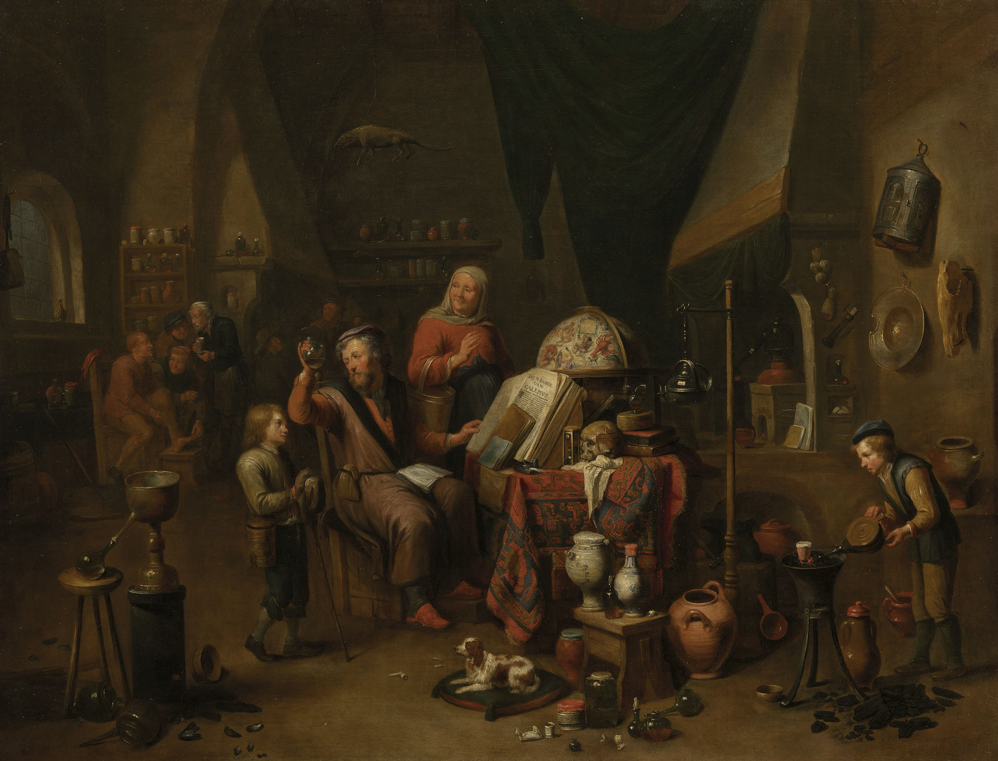
Stories of Medics and Medicine in the Royal Collection
The Royal Collection contains fascinating objects and paintings that illustrate the changing perception and use of medicine over time
LEONARDO DA VINCI (VINCI 1452-AMBOISE 1519)
The bones, muscles and tendons of the hand
c.1510-11RCIN 919009
Recto: five studies of a right hand, showing bones and muscles; six smaller drawings of digits; notes on the drawings. Verso: studies of the skeleton of a right hand; anatomical studies of fingers; studies of the skeleton of a right hand; a clenched fist; notes on the drawings.
Leonardo demonstrates the structure of the hand, building it up in the manner of an engineer. He begins at lower left with the bones, then adds the deep muscles of the palm and wrist at lower right, the first layer of flexor tendons at upper left, and the second layer of tendons at upper right. Leonardo was intrigued that the two sets of tendons bend the fingers in different ways, either with the finger held straight or curling the finger, as shown at bottom right; in each finger one tendon passes through a gap in the other, illustrated at upper centre, as he had observed in the bear’s foot more than twenty years earlier.
In the winter of 1510-11 Leonardo was apparently working in the medical school of the University of Pavia, alongside the professor of anatomy Marcantonio della Torre. His investigations focussed on the mechanisms of the bones and muscles, and he developed novel illustrative techniques to convey the complexity of these mobile, layered, three-dimensional structures.
Text adapted from Leonardo da Vinci: A life in drawing, London, 2018


